
Enhancing regional anaesthesia training with handheld ultrasound: the bridge between theory and practice
Author: Dr Gabrielle Grounds, ST7 Anaesthetics, Kent, Surrey & Sussex School of Anaesthetics
The integration of ultrasound technology into anaesthetic practice has transformed how we provide patient care. Over the past decade, handheld portable ultrasound devices have gained popularity, offering real-time imaging at the point of care.
Traditional ultrasound machines are often large, stationary devices that are restricted to individual departments, which limits their accessibility for educational and training purposes. Handheld ultrasound provides a solution, offering a compact wireless device that connects to mobile devices or tablets and delivers high-quality images through an intuitive interface. These devices facilitate ultrasound scanning in remote locations outside the clinical area, enhancing the flexibility and reach of ultrasound training and practice.
Current evidence
A review of the existing literature shows a growing body of research focused on the clinical application of handheld ultrasound at the point of care. There is a degree of reluctance to integrate handheld ultrasound devices into routine clinical practice. This hesitancy is primarily due to concerns regarding the regulation, encryption, and data protection of images captured on mobile devices or tablets connected to the ultrasound machines. While most of the existing literature focuses on the clinical application of handheld ultrasound, there has been limited attention given to its potential as an educational tool. It can enable learners to practise and refine their ultrasound skills before their application in clinical care. Additional research is needed to assess its effectiveness in training environments and to develop best practices for its role in medical education.
Theory and practice
Handheld ultrasound can provide a stepping stone for residents to become competent in a range of skills, from basic ultrasound scanning to advanced image acquisition. It further develops understanding of functional anatomy and appreciation of anatomical variations in a non-clinical, low-stress, and non-time-pressured setting. Enhancing skills in ultrasound-probe handling and image interpretation can improve procedural accuracy and assist in decision-making regarding needle insertion and injection site. Optimal performance of these skills may enhance both the efficacy and safety of regional anaesthesia block performance when translated into clinical practice.
Ultrasound in the curriculum
Regional anaesthesia is a key learning objective in RCoA’s Anaesthetic Curriculum 2021. It focuses on obtaining the competency to achieve optimal ultrasound imaging and perform ultrasound-guided regional anaesthesia for procedures involving upper and lower limbs, chest, and abdominal wall. The development of competence in ultrasound scanning, image interpretation and the recognition of anatomical variations is contingent upon the resident’s exposure to a range of regional anaesthesia blocks throughout their training, which may not always be assured. Repeated practice can help to solidify the association between sonoanatomy, prosection anatomy, and textbook anatomy.
Handheld ultrasound in practice
In our department, we examined the potential benefits of offering anaesthetic residents, on a six-month rotation, a handheld ultrasound device for voluntary, self-directed learning. Our goal was to enhance their reported confidence in regional anaesthesia through improved ultrasound technique, image acquisition, and anatomical interpretation. A business case was submitted to the local trust for funding through the education budget to procure the GE Vscan Air™ handheld ultrasound for the anaesthetic department. The funding was granted and the device purchased by the trust with the stipulation that the handheld ultrasound be utilised exclusively for training purposes and not for clinical application.
A real-time demonstration of the device was provided, and residents were given a reference handbook to facilitate its use. Anaesthetic registrars were able to borrow the handheld ultrasound on a rotational basis, allowing them to take the device home for non-clinical practice during their personal time. Qualitative feedback was collected before and after the use of the device.
The handheld ultrasound was well received, with six out of the seven residents voluntarily borrowing and utilising the device in the non-clinical setting. Analysis of the feedback from these residents revealed that 83% reported an improvement in their ultrasound scanning technique and 100% recognised an improvement in their ultrasound-image interpretation skills. All residents surveyed would recommend the handheld ultrasound as an educational tool for developing regional anaesthesia skills in anaesthetists in training.
Handheld ultrasound may serve as a valuable educational tool for anaesthetic residents, providing a bridge between theoretical regional anaesthesia study and real-time clinical practice. By enabling frequent, flexible and low-pressure learning, it allows residents to independently practise ultrasound scanning, thereby improving proficiency in image acquisition and sonoanatomy interpretation. Ultimately, this can lead to greater self-confidence in ultrasound probe handling for performing regional anaesthesia and facilitate attainment of the competencies outlined in the RCoA Anaesthesia Curriculum 2021. We encourage anaesthetic departments to acquire similar devices for educational purposes through their respective education departments.
Further research and validation studies are required to fully understand the educational benefits of handheld ultrasound. It is vital that we evaluate the cost-effectiveness and broader implications of handheld ultrasound in terms of accessibility, training outcomes, and impact on clinical practice.
Figure 1
Schematic representation of median nerve anatomy and related structures superior to the elbow crease
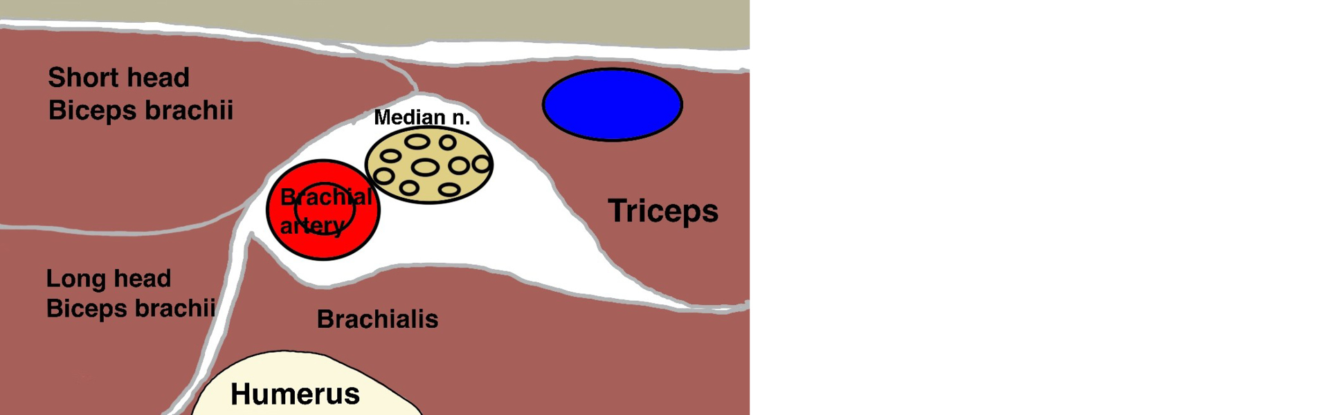
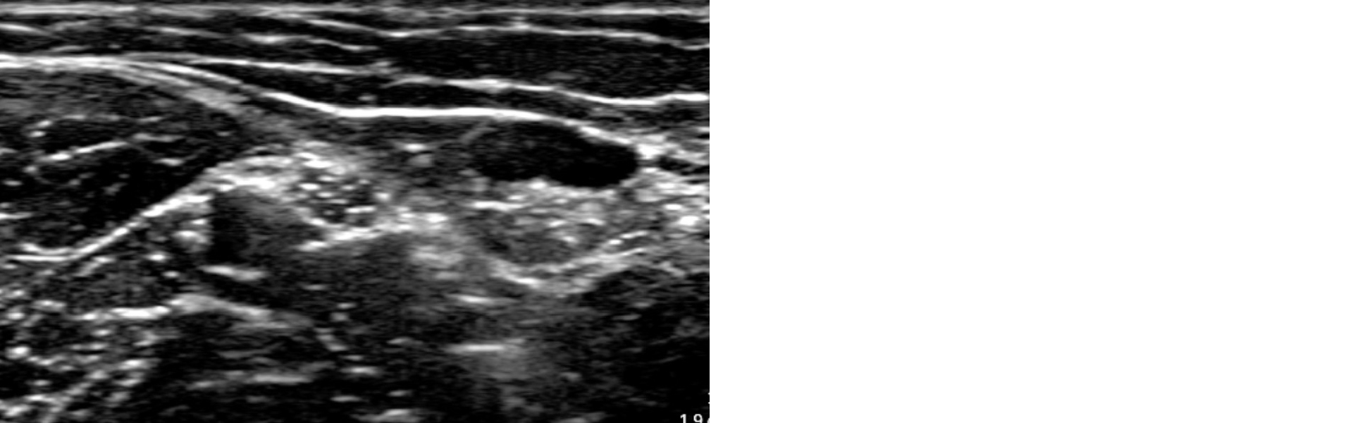
Figure 2
Ultrasound image of the median nerve superior to the elbow crease in the upper limb obtained using the handheld ultrasound device GE Vscan AIR™ CL
Acknowledgments
Thank you to Dr Benjamin Carey, Consultant Anaesthetist, Queen Victoria Hospital, East Grinstead, who supported the procurement of the handheld ultrasound device and resident education in its use.